A new radio-imaging, diagnostic agent to guide targeted therapy for epithelial ovarian cancer
Overview
Professor Hooper’s team are testing an innovative formula, developed using a small, safe dose of radioactive particles, which could elevate scanning techniques for more effective ovarian cancer diagnosis and targeted treatment.
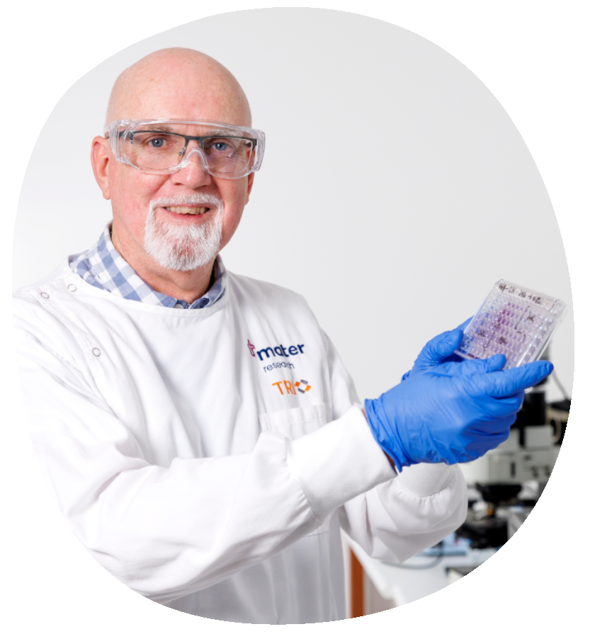
Lead researcher: Professor John Hooper
Grant received: $60,000 for 5 years contributed by the OCRF to a Medical Research Future Fund grant totalling $1.25M
OCRF research pillar: Early Detection, Treatment
Primary institution: Mater Research Institute
Latest update
We’re proud to have a phase 1 clinical trial currently underway with recruitment via Mater Hospital and the Royal Brisbane Hospital. We’re testing this new approach for diagnostics and treatment of ovarian cancer, whereby we administer our new agent which attaches a tracer to the ovarian cancer cells. This is expected to make detection of the disease easier during scans, similar to what is currently performed with prostate cancer diagnosis.”
Professor John Hooper, August 2025
Project details
Professor John Hooper and his team are investigating improving ovarian cancer outcomes with a theranostics approach — precision medicine that combines accurate diagnostics and targeted treatment into the same approach.
The imaging formula they are testing uses safe doses of radioactive particles that could improve the effectiveness of Positron Emission Tomography (PET) scanning techniques, better displaying cancer presence and location to enable targeted treatments, or assist surgeons to visualise cancer prior to operations.
Professor Hooper has been researching this approach for over two decades. The spark began during his postdoctoral studies in the United States where the lab he was working in had identified a series of antibodies — tiny molecules that were binding to proteins on the surface of cancer cells and helping them to grow. If they could work out what those antibodies were actually binding to on the cancer cell surface, maybe they could stop this binding from occurring, and subsequently stop the cancer progressing.
His colleagues didn’t know how to confirm what on the surface of cells enabled the antibody binding. However, thanks to his experience in innovative bioinformatics and proteomics techniques, Professor Hooper did. He identified a protein called CDCP1 on the cancer cell surface, which plays a role in cancer growth and spread. He then focused on developing a second antibody that was able to stop CDCP1 — called 10D7.
With OCRF and Medical Research Future Fund funding, the team are testing their new radio-imaging agent to see how effectively it firstly allows them to visualise specific ovarian cancer cells in preclinical models, then, if cancer presence is confirmed, test the effectiveness of an injected, second formula designed for treatment.
Aims:
The team aim to:
- Test their formula for diagnostics and therapy in preclinical models, including testing a library of novel theranostic formulas for epithelial ovarian cancers, including high-grade serous ovarian cancer which is the most common and aggressive subtype.
- Validate the performance and safety of the formulas, including the radioactive particles involved, and prepare it for a first-in-human study.
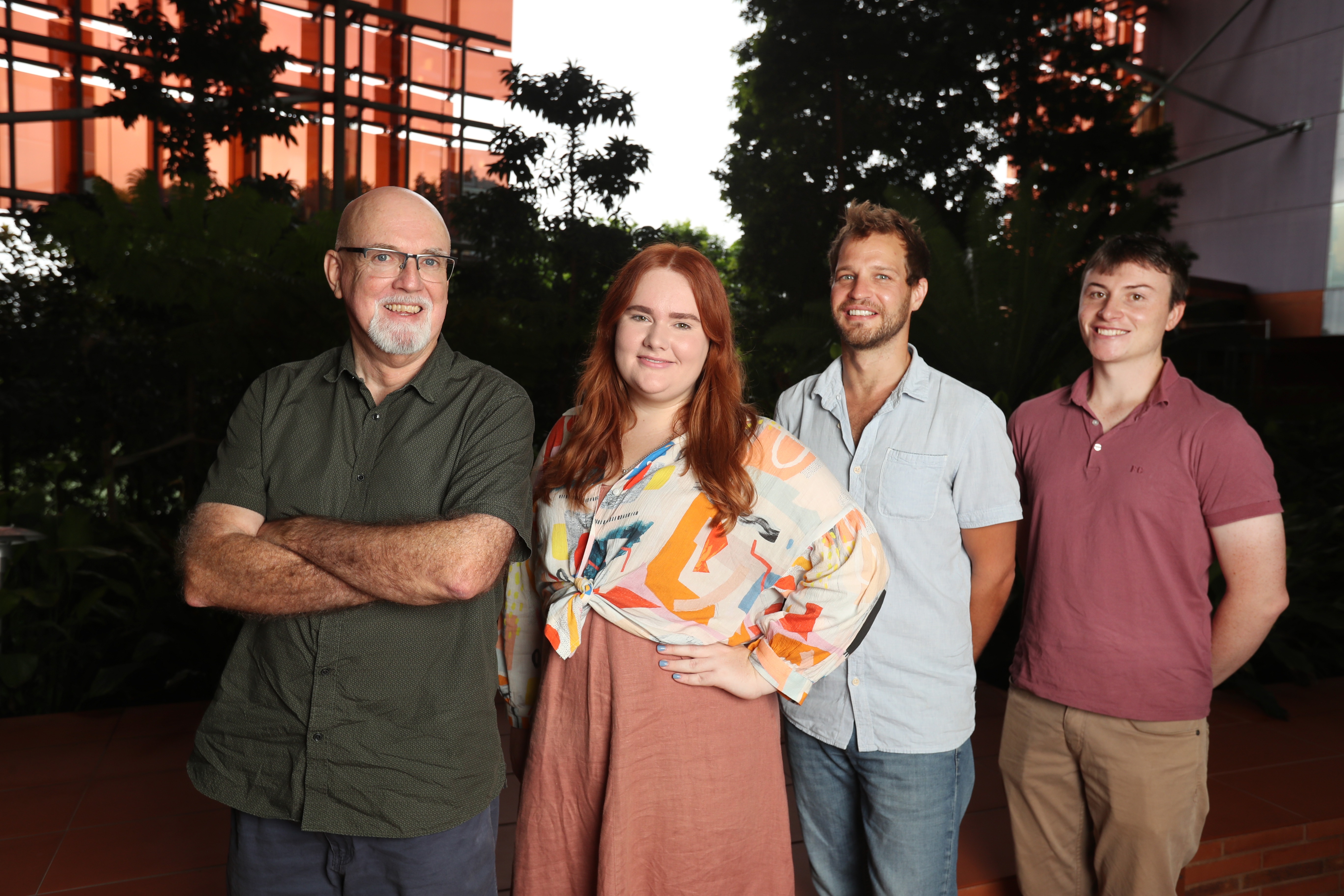
(Pictured above: Professor John Hooper and his team)
Approach:
First the team need to identify how commonly the CDCP1 protein is present on patient tumours, using immunohistochemistry techniques, to confirm whether targeting it will stop cancer in many patients.
Secondly, they need to test, in ovarian cancer cell lines, how well the antibody can directly target CDCP1, and also test how strongly it binds to the CDCP1 on the cancer cells. This will help them understand whether the treatment can selectively target ovarian cancer cells, or if it is also targeting healthy cells which can lead to side effects.
On a molecular level, the diagnostic formula works like this:
- CDCP1 is the protein on the surface of the cancer cells.
- 10D7 is the antibody Professor Hooper identified that can stop the cancer-causing functions of CDCP1.
- Zirconium-89 is the radioactive particle attached to 10D7, which then binds to CDCP1, allowing PET scans to visualise the cancer.
The team will engineer the 10D7 antibody, attaching it to the Zirconium particles, so that 10D7 ‘docks’ or binds to the Zirconium. The engineered particles will then be injected into sophisticated preclinical models that mimic a human tumour environment, moving through blood circulation until they find cancer cells with the CDCP1 protein on the surface. The Zirconium particle then gives off a signal that’s detected by a PET scanner, allowing clinicians to understand if cancer is present, and if so, where it has spread to.
The team are also confirming that the radioactive particle Zirconium-89 only emits a safe amount of energy so that the PET scanner can detect it while ensuring safety for human use.
Ambition and outcomes:
Twenty years after first identifying CDCP1 on cancer cells, Professor Hooper’s research, and the results of this project have paved the way for a phase 1 clinical trial that is currently underway.
Promising results of this project include:
- The team finding that in around 80% of the diverse ovarian cancer subtypes they reviewed, CDCP1 was present at high levels, with much fewer healthy cells affected, suggesting that a treatment targeting this protein could benefit many patients with minimal side effects. They also found that CDCP1 is present on aggressive prostate, breast, pancreatic and bladder cancers, so this work could lead to improve diagnostics and treatments across cancer types.
- When they tested the treatment formula, the results were so good that the team could barely find the remnants of the tumour when they went back to inspect the preclinical models.
The phase 1 trial will involve approximately 12-30 patients who have ovarian cancer and will allow the research team to confirm the agent’s diagnostic effectiveness in humans.
If their first trial is successful, and identifies a patient with strong accumulation of the theranostic agent which would indicate ovarian cancer and where it has metastasised, they then hope to test the ability of the same approach as a targeted treatment. This would be done by switching out the Zirconium-89 for a higher energy radioactive particle, LeticiumU-177, which would eliminate the cancer cells it has attached to.
To prevent significant side effects, the formula would also involve a method to block the healthy cells that also have CDCP1 on their surface—leaving only the ovarian cancer cells exposed to the radioactive particle treatment.
With ovarian cancer metastasis and recurrence being persistent and urgent issues, Professor Hooper’s approach hopes to provides accurate visualisation of cancer cells, better information for treating clinicians and a new treatment option for advanced ovarian cancer — that could significantly improve outcomes for patients.
Current status:

For every project like this, many more can’t get underway due to a lack of funding. Support research like this to help them move forward.
Share page

Get the latest news, stories & updates.
