Discovery of new sugar-based markers in blood and using magnetic resonance spectroscopy towards early ovarian cancer detection
Overview
This project will investigate how effective specific sugar-based biomarkers and innovative MRS scanning techniques are at detecting ovarian cancer in the earliest stages.
Lead researcher: Professor Michael Jennings
Grant received: $1,403,190 for three years
OCRF research pillar: Early Detection
Primary institution: Griffith University
Associated institution/s: Princess Alexandra Hospital and Mater Hospital Brisbane
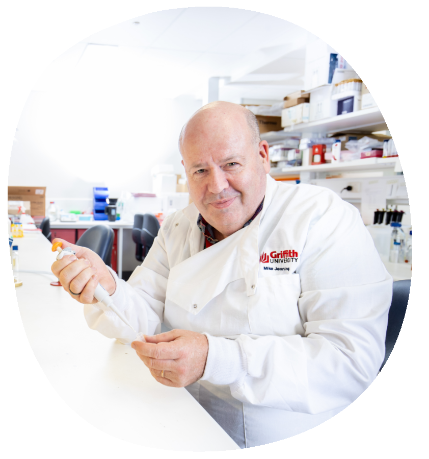
Last year we secured multi-institutional agreements, hired required staff in the lab and multiple research nurses at both hospital sites. We hired a scientist who is helping with sample collections in the clinic and in the lab including a mass spectrometry expert working up the approaches we will now take with our samples. With secured ethics, we have begun our sample collections.”
Professor Michael Jennings, June 2024
Project details
This project combines the talents of two world-leading researchers: Professor Carolyn Mountford who has developed innovative magnetic resonance spectroscopy (MRS) technology and Professor Michael Jennings who is an expert in glycomics.
Glycomics and ovarian cancer early detection
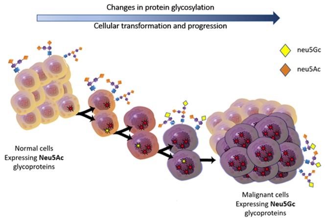
Glycosylation on cells involves carbohydrate sugars, including those that attach to proteins, which can help cell function but can also be involved in a range of diseases. These are different to the sugars we eat, which are mainly used as energy sources or building blocks for various bodily functions.
Professor Michael Jennings is determining whether one specific sugar, n-glycolyneuraminic acid (Neu5Gc), could act as an ovarian cancer early detection biomarker.
The team discovered that Neu5Gc is present in the blood of healthy people at very low levels, but its level increases if cancer is present. With OCRF funding they are investigating how well it indicates ovarian cancer at each stage, including the very earliest stages, to determine if it could provide the foundations of an effective early detection test.
Neu5Gc links itself to CA-125 proteins. CA-125 is currently used to monitor ovarian cancer remission and recurrence, as it can be elevated if ovarian cancer is present, but it can also be elevated due to other non-cancerous conditions such as endometriosis. This makes it unreliable for ovarian cancer early detection on its own. However, in previous studies the team have found that Neu5Gc only links to the cancerous CA-125 proteins – so it could be the key to a stand-alone ovarian cancer test, or to enhancing the current CA-125 test.
Non-invasive MRI and MRS scanning for early ovarian cancer
Traditional Magnetic Resonance Imaging (MRI) is a non-invasive imaging technology that produces three dimensional detailed anatomical images. In this project MRI will be used to locate an ovarian lesion, before using MRS to evaluate the tissue chemistry. Professor Mountford has pioneered the science behind monitoring tumour development and progression with these techniques. The resultant chemical data is fed into a cloud-based system developed by Professor Mountford’s team at DatChem, which then returns a cancer risk assessment to the radiologist. The question at hand is—can the MRS method detect very early small cancers in the ovaries of women at high risk earlier than current imaging methods?
In a parallel program the MRS technique identified, on blood specimens from women with ovarian cancer, fucosylated glycans shed from the tumour and attached to a particular serum called Lipoprotein ‘a’. This project will confirm earlier studies of levels of lipoprotein (a) in the blood of ovarian cancer samples and seek to identify the specific types of fucosylated glycans shed from the tumour.
Aims:
Professor Jennings team aims to validate the effectiveness of Neu5Gc as a biomarker by:
- Collecting samples and establishing a dataset from four groups of patients: those with suspected ovarian cancer who have had symptoms and a CT scan but haven’t been biopsied, those at familial risk of ovarian cancer, those with BRCA gene mutations, and healthy control samples.
- Sampling from diverse locations including urine, cervical smears, and blood, to evaluate which bodily fluid is most effective for use in detecting Neu5Gc.
- Analysing samples to evaluate the effectiveness of Neu5Gc as a biomarker by itself and how well it indicates various stages of ovarian cancer.
- Evaluating the effectiveness of Neu5Gc combined with CA-125 to see if it enhances accuracy.
Additionally, the team will:
- Determine whether the Neu5Gc biomarker can lead to discovery of additional ovarian cancer biomarkers associated with CA-125.
- Identify, at the cellular level, what specifically elevates Neu5Gc levels in ovarian cancer. Neu5Gc is also elevated in the blood of people with melanoma and breast cancer, so the team need to differentiate between these diseases to create an ovarian cancer-specific test.
Meanwhile, Professor Mountford’s team will use MRI and MRS technology to:
- Determine whether the MRS technology can identify early and small ovarian cancer and confirm the presence of small tumours with levels of Neu5Gc. They will also correlate these results with the risk assessment returned via the MRS scan and DatChem system.
- Develop the MRS technology so that it is sensitive enough to get as much information from an ovary scan as they currently can get from a breast scan and provide an accurate risk assessment based on a risk score spectrum. The aim is to determine how early the MRS technology can identify ovarian cancer.
Approach:
Professor Mountford’s work with breast cancer has informed their new approach to ovarian cancer scanning. In breasts, this technique can identify the transition from healthy tissue to intermediate states and even tiny foci, showing microscopic images of tumour cells forming. Undertaking the same 2D MR technology on the smaller ovary is quite challenging but early results are promising. Moreover, the 2D data from the ovary provides information on the extent of transition from normal to cancerous and, if cancerous, the cellular differentiation. Professor Mountford’s team will:
- Use 3TMRI scanners to preoperatively report on an ovarian lesion, or apparently healthy ovary, and provide a chemical analysis and risk assessment. To assess the ovarian cancer the surgeon will then take a biopsy during surgery and confirm whether Professor Mountford’s original report gave accurate results. For the risk prediction, a longitudinal study will be undertaken, similar to the breast study now completed.
- With a cohort of patients who have had breast cancer and are at high risk of ovarian cancer, conduct an MRS 3T examination of their ovaries to identify any transition towards a cancer or small foci of cancer at a time when surgery or therapy could be effective.
Invention of SubB2M
Collaborating with hospitals, Professor Jennings’ team will build their own sample collection and clinical database and establish a collection pipeline.
By including at-risk and suspected ovarian cancer patients in the study, the likelihood that they will discover a patient has early-stage ovarian cancer is increased by testing the Neu5Gc. This means that as well as benefitting the patient, they can collect rare early-stage samples.
With researchers from the University of Adelaide, Professor Jennings’ team invented a tool called SubB2M, which was developed to detect specific sugars associated with cancer. The team engineered this SubB2M tool to specifically attach to and detect Neu5Gc. This invention allows the team to measure the level of Neu5Gc in bodily fluids, to specifically detect ovarian cancer and indicate its stage.
When blood (serum) samples are collected from people with ovarian cancer, the team will use their SubB2M tool to capture every molecule in the serum that contains Neu5Gc therefore capturing only cancer-associated sugars. Everything else is washed away, leaving only CA-125 proteins that have Neu5Gc sugars attached and potentially other ovarian cancer biomarkers.
Ambition and outcomes:
This project could validate Neu5Gc as an effective biomarker for ovarian cancer and enhance the accuracy of CA-125 testing. Furthermore, it could uncover unknown biomarkers for ovarian cancer. Short-term, the project will benefit those participating in the study by uncovering if any of the donors have high levels of Neu5Gc and therefore likely have cancer, giving these patients the opportunity to be treated earlier.
Professor Mountford’s scanning technique could empower patients to make informed decisions about both their risk of ovarian cancer and their ovarian cancer treatment, as well as better informing clinicians before surgery. Additionally, the results of this study will contribute to Food and Drug Administration (FDA) approval for use of Siemen’s 3T scanners in ovarian cancer risk assessment and diagnostics. In approximately five years Professor Mountford’s team hope to have this technology in use in several sites across Australia for those at high risk.
According to Professor Jennings, multi-year OCRF funding affords the focus and coordination to simultaneously collect and analyse samples, condensing the project timeframe from approximately nine to three years — delivering answers faster.
Longer-term this project is key to establishing a glycomics-based approach as a new method for ovarian cancer early detection, which could mean it is caught at the earliest stages when it is most treatable, improving a patient’s chance of survival. It could also establish MRS scanning as a non-invasive early detection technique, to better informing patients and clinicians earlier.
Current status:
This team preoperatively evaluates the chemistry of the ovarian lesion whereupon a research report is generated to predict if the lesion is malignant or benign. This capability has been in place since 2006. The researchers are conducting extensive studies in the lab with samples to determine if a blood test for ovarian cancer is predictive whereupon an MRI and MRS scan can be undertaken to verify the result and determine if MRI/MRS technology is effective and correct. The challenge is to know who needs a scan and the blood test will provide this information *
*Want to learn more about the medical research pipeline? Read more here.

For every project like this, many more can’t get underway due to a lack of funding. Support research like this to help them move forward.
Share page

Get the latest news, stories & updates.
